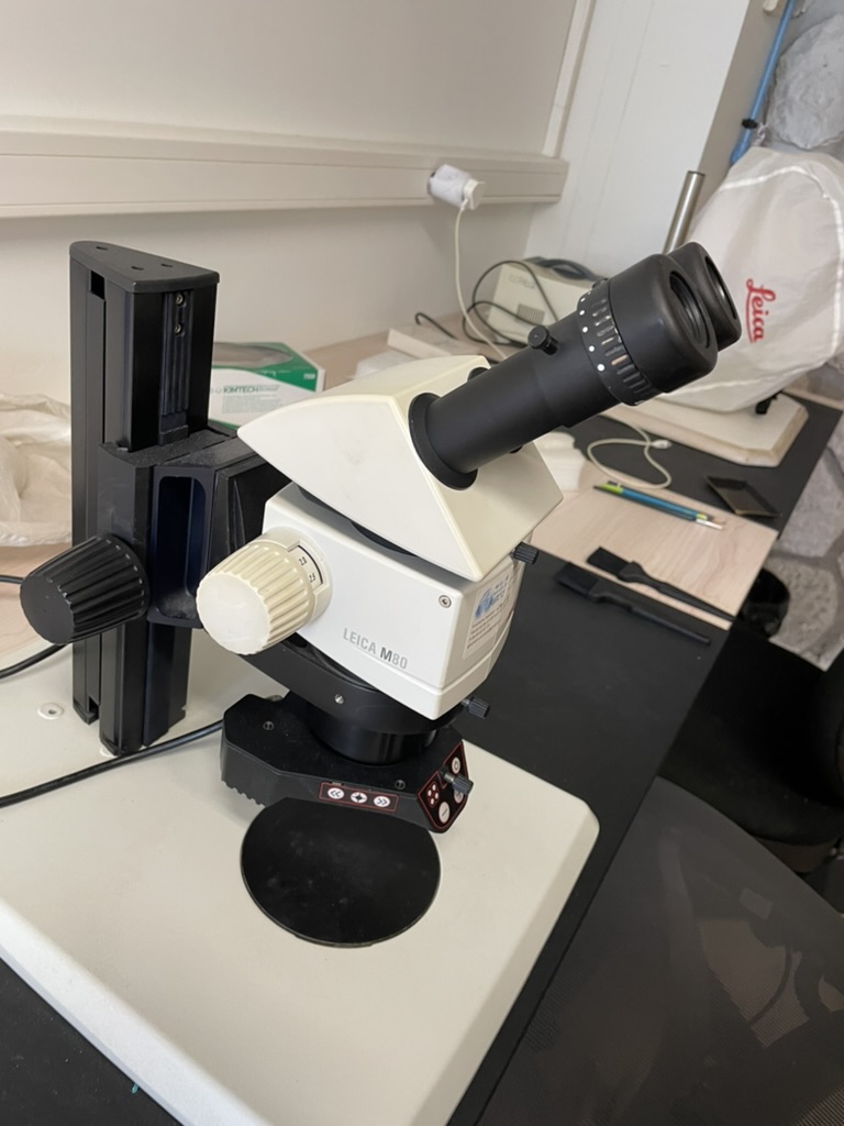
The MANTA platform manages a park of manual and automated stereomicroscopes, geared with some cameras, in open access at CEREGE, in room 109.
Book your slot here : https://grr.osupytheas.fr/day.php?year=2025&month=04&day=17&area=150
Microfossil Analyses: New Technologies and Applications
The MANTA platform manages a park of manual and automated stereomicroscopes, geared with some cameras, in open access at CEREGE, in room 109.
Book your slot here : https://grr.osupytheas.fr/day.php?year=2025&month=04&day=17&area=150
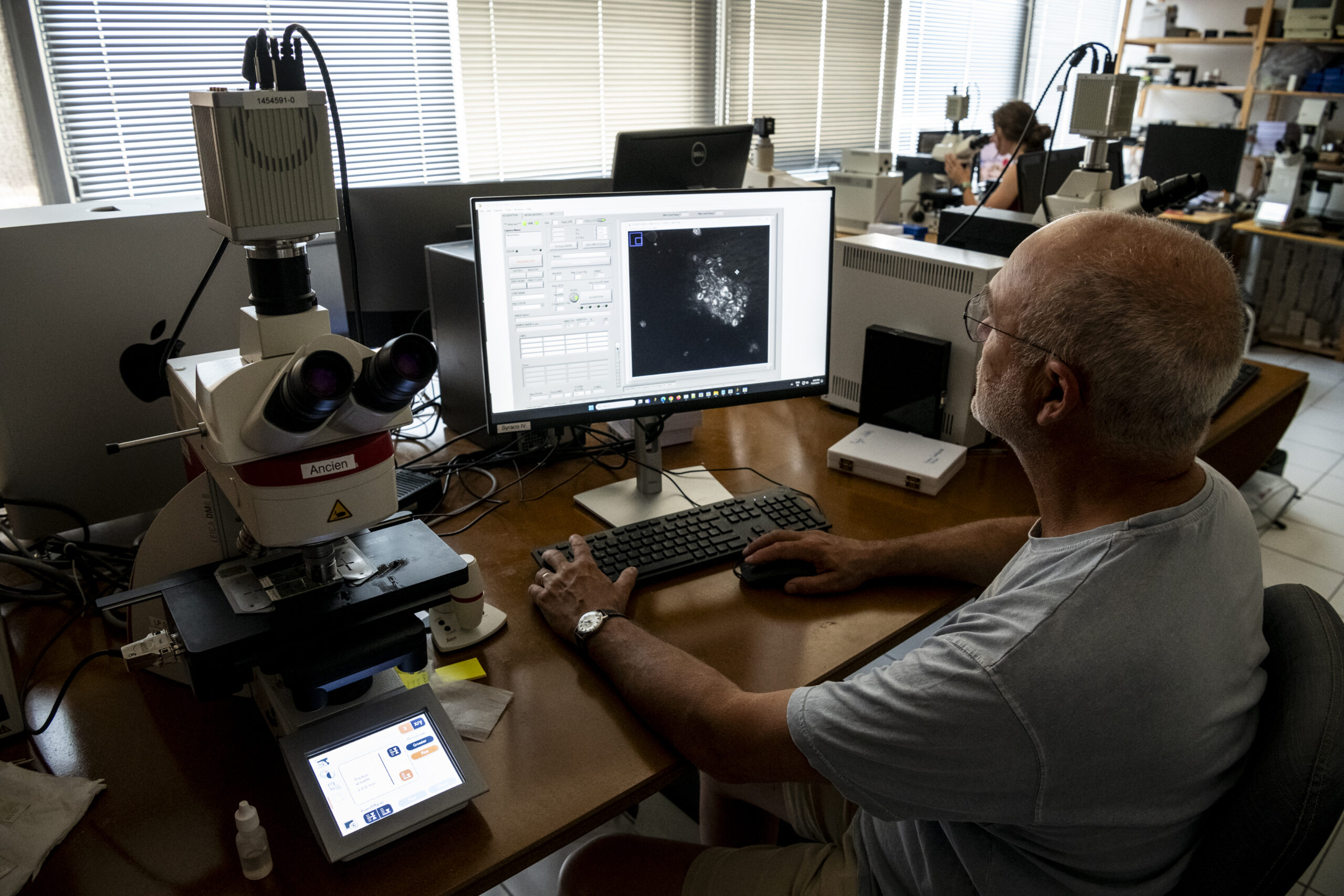
The MANTA platform hosts 3 Leica DM6000 microscope, with a x100 objective having a numerical aperture of 1.47, and a condenser lens having a 1.2 numerical aperture.
Avaliable filters :

Three circular polarizers made by Chroma Technology Corp. are integrated in the microscope. One right circular polarizer is positioned as analyzer. It consists of a linear polarizer oriented at +90° placed below a 1/4 lambda plate oriented at +45° mounted in a Leica cube and placed in the upper automatic turret of the microscope. This is a convenient place when one wants to automatically remove this analyzer to use other filters. If this not necessary, the analyzer can be placed in its regular position.
One (and one) left (right) circular polarizer, LCP (RCP), made of a 1⁄4 lambda plate oriented at 45° (-45°) followed by a linear polarizer oriented at 0°. These two polarizers are used alternatively when taking images of the same crystal. LCP and RCP are placed in the revolving filter chamber of the automated condenser block. For manual use, a 1⁄4 lambda plate could be placed under a linear polarizer, and rotated manually from -45° (LCP) to 45° (RCP).
One of five monochromatic bandpass filters centered at 435, 460, 560, 655, and 700 nm (AT435/20X, AT460/50M, ZET561/10X, AT655/30M and ET700/50M; all from Chroma Technology Corp.) is positioned in the light trajectory after the light bulb. The 561 nm filter is used in routine work because of its versatility (see below). The other filters are used to test the method and in special occasions when studying relatively thick calcite particles in the range of 1.4-1.9 μm in the case of 700 nm; or for detailed measurements of thin particles in the range of 0.2-0.4 nm for the 435 nm filter.

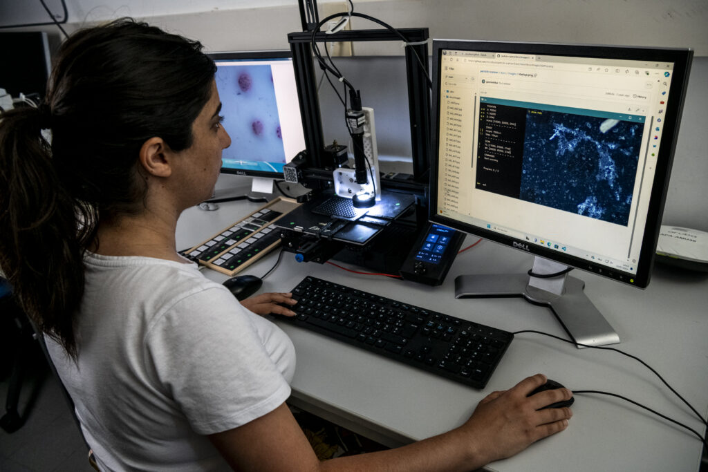
“SASHIMI (System for Automated Scanning and High-resolution Imaging of MIcrofossils) is a low-cost device designed to capture 2D images of microfossils and particles. It is equipped with a Basler camera mounted on a motorized XY stage. This system enables Z-stack imaging of particles, with a resolution of ~5 µm.
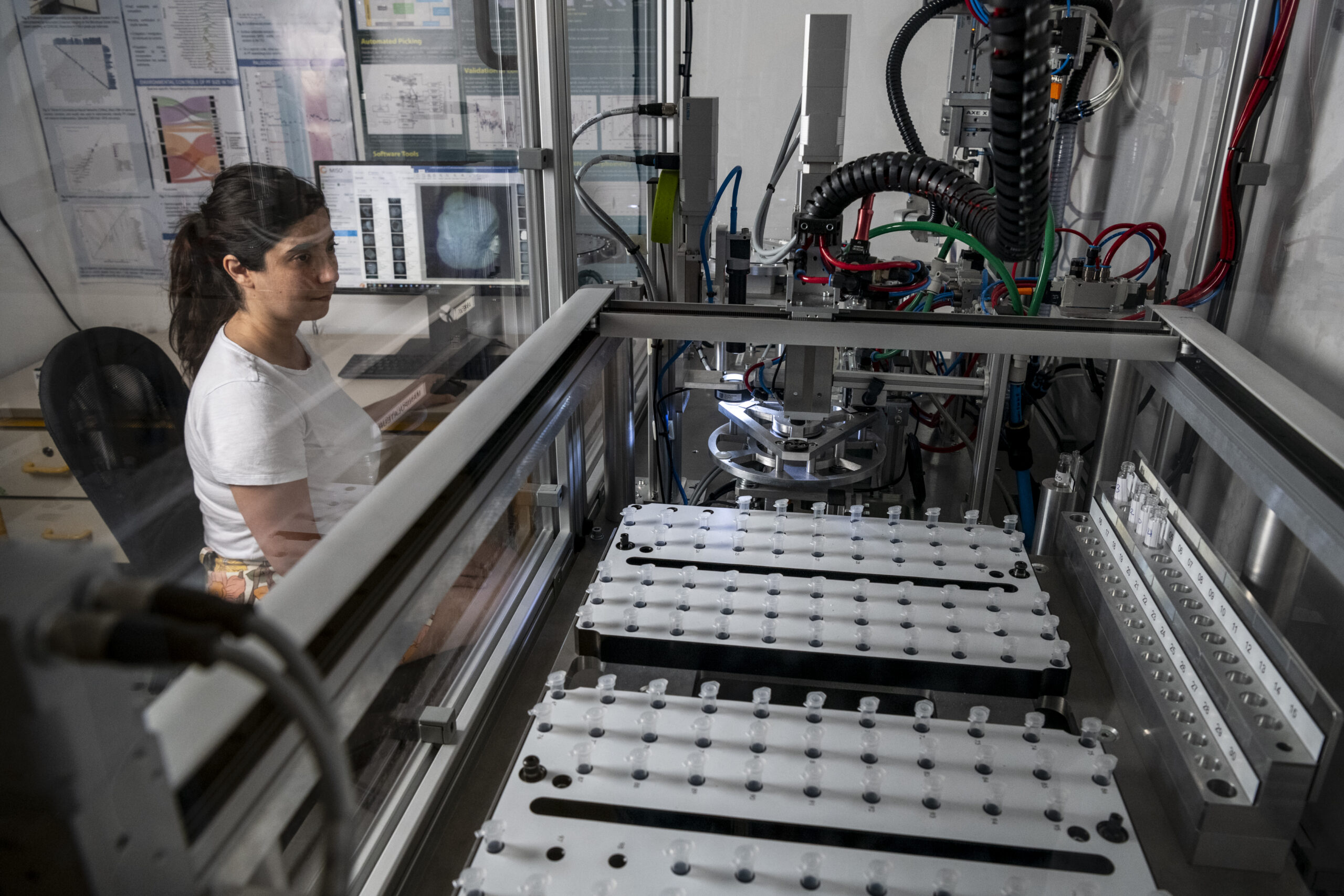

The automatons MiSo (Microfossil Sorter) are fully controlled by a dedicated software which handles the control of the automaton (set-up, calibration, troubleshooting), management of the sample processing (carrousel description, metadata input, sorting rules by size and neural networks outputs), control of camera settings, image processing (segmentation, collation, depth-stacking, background subtraction), CNN models handling (pb, onnx), live vizualisation, error controlling. This software is licensed to ATG Technologies and available with new MiSo machines. Current version is 3.1, and works on Windows OS.
MiSo v1 : the first generation of the Microfossil Sorter. System patented https://patents.google.com/patent/FR3122586B1/fr
MiSo v2 : the second generation of the Microparticle Sorter : includes a double imaging system (with 2 cameras and attached lighting and optical tubes on double cups) ; an automated cleaning system of the channelling system.